Bio-degradable cards
Every card imaginable!
|
Bio-degradable cards Every card imaginable! Can I Have an MRI with a Metal Implant? Your Complete Guide to Safety and ProtocolsFacing an MRI scan can be a little daunting on its own. The loud noises, the enclosed space, the need to stay perfectly still – it’s a unique experience. But when you have a metal implant, a whole new layer of questions and anxieties can surface. Is it safe? Will it hurt? Could the scan even work? If these questions are on your mind, please know that you're not alone and your concerns are completely valid. The good news is that having a metal implant doesn't automatically mean you can't have an MRI. Millions of people with implants undergo these scans safely every year. The key to ensuring your safety lies in understanding the process, knowing your implant, and communicating clearly with your medical team. This guide is here to walk you through everything you need to know. We’ll break down the science in simple terms, explain the different safety classifications for implants, and give you the confidence to navigate your MRI appointment. Our goal is to replace your worry with knowledge, so you can focus on what’s most important – your health. Table of Contents
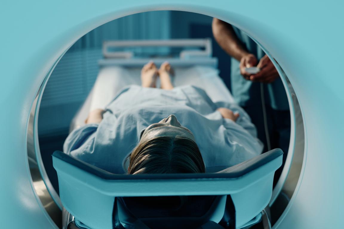
Why MRIs and Metal Can Be a Risky Mix: Understanding the ScienceTo understand why your medical team takes metal so seriously, it helps to know a little bit about how an MRI scanner works. Don't worry, we won't get too technical. Think of an MRI machine as a giant, incredibly powerful magnet. It's thousands of times stronger than a typical fridge magnet. Its job is to create a strong, stable magnetic field around your body. When you're inside the scanner, it also sends out radiofrequency waves. These waves interact with the protons in your body's water molecules, temporarily knocking them out of alignment. When the radio waves are turned off, the protons relax back into place, releasing energy signals. A computer then cleverly interprets these signals to create incredibly detailed, cross-sectional images of your organs, soft tissues, and bones. This amazing technology is what allows doctors to see what's happening inside you without making a single incision. However, when you introduce metal into this powerful magnetic environment, three potential problems can arise.
1. The Magnetic Pull: Force and TorqueThis is often the first thing people worry about. If an implant contains magnetic metals, like iron or certain types of steel (known as ferromagnetic metals), the MRI's powerful magnet can pull on it. This can cause two types of movement:
It's important to know that most modern medical implants, like joint replacements made from titanium or cobalt-chromium alloys, are not strongly magnetic and are designed with MRI safety in mind. However, older implants, surgical clips, or fragments of metal from a past injury could be ferromagnetic, making this a critical safety check.
2. Radiofrequency (RF) HeatingThe second major concern is heating. During the scan, the radiofrequency waves we mentioned earlier can be absorbed by metallic objects. This can cause the implant to heat up significantly. Think of it like how metal can get dangerously hot in a microwave oven, although the scientific principle is different. This heating can be intense enough to cause thermal injury or burns to the delicate tissues surrounding the implant. The amount of heating depends on the type of metal, as well as the shape and size of the implant. A long, thin wire, for example, can sometimes act like an antenna, absorbing more energy and becoming hotter than a solid, compact implant. Radiographers are trained to use specific scan settings to minimise this risk for patients with "MRI Conditional" implants.
3. Image Distortion: The Artefact ProblemThe third issue isn't a direct danger to your body, but it can make the MRI scan useless. The presence of metal can severely distort the magnetic field in the area around it. This scrambles the signals that the scanner is trying to read, creating large black spots, warped areas, or bright, strange shapes on the final images. This is known as a "metal artefact." If the implant is located in or near the area your doctor needs to examine, this artefact can completely obscure the view. For example, if you have a hip replacement and your doctor needs to look for a problem in your pelvis, the artefact from the implant might hide the very thing they are trying to see. This can lead to a failed scan and an inconclusive result, meaning you've gone through the whole process for nothing. Understanding these three risks – the pull, the heat, and the distortion – is why the pre-scan safety procedure is so thorough. It’s a carefully designed process to ensure the benefits of the MRI far outweigh any potential risks. 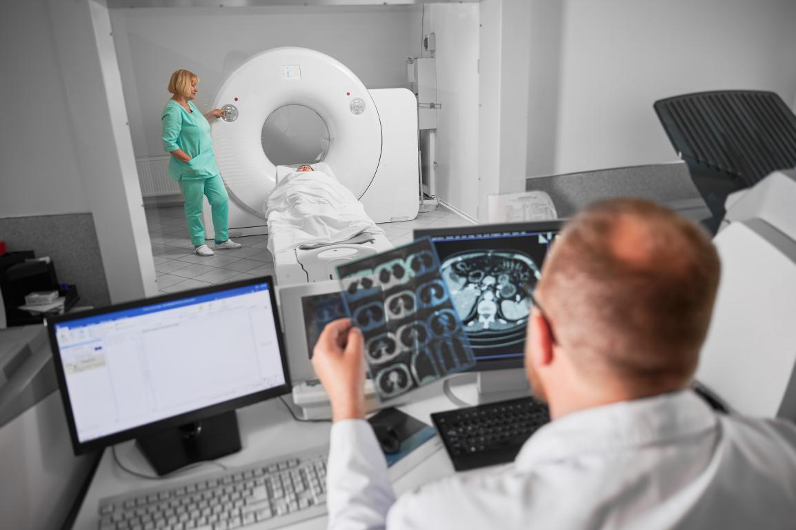
"MRI Safe," "MRI Conditional," and "MRI Unsafe": What Do They Really Mean?When you tell your medical team you have an implant, they will need to determine its MRI safety status. You'll hear them use specific terms: "MRI Safe," "MRI Conditional," or "MRI Unsafe." These aren't just casual descriptions; they are official classifications with very precise meanings. Let's break down what each one means for you.
MRI UnsafeThis is the most straightforward category. An "MRI Unsafe" device is one that is known to pose a clear and significant risk to a patient in an MRI environment. These implants are considered an absolute contraindication, meaning you should not have an MRI scan under any circumstances.
If you have an "MRI Unsafe" implant, your doctor will not proceed with the scan and will work with you to find an alternative way to get the images they need.
MRI SafeThis is the ideal, but less common, classification for implants. An "MRI Safe" device is one that poses no known hazards in any MRI environment.
If your implant is officially labelled "MRI Safe," you can undergo a scan without any special precautions related to the implant itself. However, it's still absolutely vital to declare it, as the medical team needs to be the one to confirm its "Safe" status.
MRI ConditionalThis is by far the most common category for modern medical implants, and it's also the most complex. "MRI Conditional" means the implant is safe for an MRI, but only under a specific set of conditions. If those conditions are not met, the scan could be unsafe. Think of it like a set of driving instructions. Your car is safe to drive, but only if you stick to the speed limit, stay on the right side of the road, and follow the traffic lights. For an "MRI Conditional" implant, the radiographers have a similar set of rules they must follow to ensure your safety. These "conditions" are very specific and can include:
Why the Exact Model Number is CrucialFor an "MRI Conditional" device, the MRI team cannot proceed without knowing the exact manufacturer and model name or number of your implant. They use this information to look up the device in a comprehensive database. This database tells them the precise conditions under which that specific model was tested and proven to be safe. Without this exact information, they cannot verify the conditions and, for your safety, will have to assume it's unsafe. They will not take a guess. This is why just knowing you have a "titanium hip" isn't enough; they need to know if it's a "Global Orthopaedics Model XYZ-123 Titanium Hip." This is the single biggest reason why keeping a detailed record of your implant is so important. It’s the key that unlocks a safe MRI scan for millions of people. 
The Golden Rule: Why You Must Always Inform Your Medical TeamIf there is one single piece of advice to take away from this guide, it's this: you must always, always inform the medical staff about your implant. This isn't just a helpful tip; it's the most critical step in ensuring your safety. Many people think, "My hip replacement is made of titanium, and I know that's not magnetic, so I don't need to mention it." This is a dangerous assumption. As we've seen, safety isn't just about magnetism. It's also about heating and image quality, and even non-magnetic implants are almost always classified as "MRI Conditional," requiring very specific protocols. Who Do You Need to Tell? You should consider it your responsibility to inform every single person involved in your care pathway. Redundancy is a good thing when it comes to safety.
Why is This Repetition So Important? It may feel like you're repeating yourself, but this system of checks and double-checks is designed to catch any potential issues. The radiographer who physically operates the scanner has the final responsibility for your safety. They will take your information, verify your implant's "conditional" status in their database, and set the scanner to the exact parameters required for your specific device. They are your ultimate safety net. By giving them clear and accurate information, you are empowering them to do their job properly and protect you from harm. Never assume that the information has been passed on from your GP. Never assume that because you mentioned it on the phone, the person on the day will know. Always take personal responsibility for communicating this vital piece of information at every step. It’s a simple action that has a profound impact on your wellbeing. 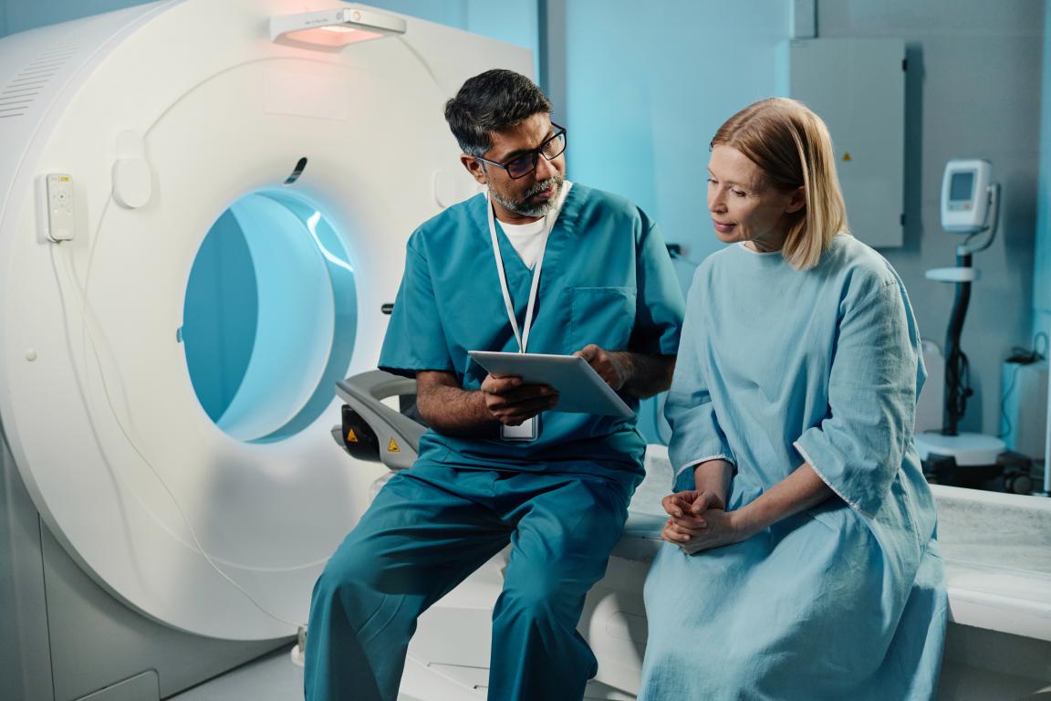
The Vital Information: What Your MRI Team Needs to Know About Your ImplantWhen you inform the MRI team about your implant, they are going to ask for some very specific details. Simply saying "I have a knee replacement" or "I have a stent" isn't enough information for them to make a safety assessment. To ensure your scan can go ahead safely and effectively, they need the precise details to look up your device in their safety database. Being prepared with this information can make the difference between a smooth appointment and a cancelled one. Here’s exactly what you need to have ready. The Four Key Pieces of Information
Without the manufacturer and model number, the team cannot look up the specific "MRI Conditional" guidelines. They will not be able to proceed and will have to cancel your scan for safety reasons. So, How Do You Find This Information? This is often the biggest challenge for patients, especially if the surgery was many years ago. It’s not information most of us carry around in our heads. Here are the best places to look:
Finding this information can sometimes take time and a few phone calls, so it's best to start this process as soon as you know you might need an MRI. Don't wait until the day before your appointment. Being proactive and organised will significantly reduce your stress and ensure the medical team has everything they need to take care of you safely. 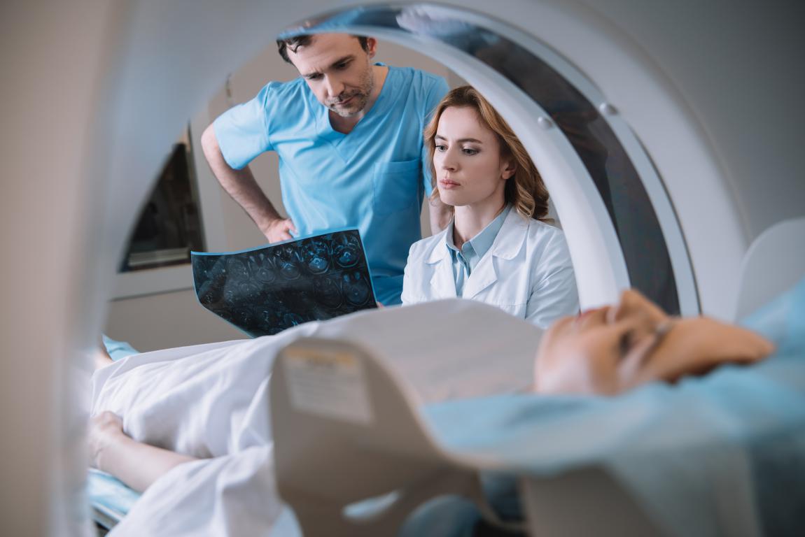
When an MRI Isn't an Option: Exploring Alternative Imaging ScansWhat happens if you have an "MRI Unsafe" implant, or if you simply cannot find the necessary documentation for your "MRI Conditional" device? First of all, don't panic. An MRI is a powerful tool, but it's not the only way for doctors to see what's going on inside your body. If an MRI is ruled out, your doctor will choose the next best imaging technique based on what they need to diagnose or investigate. Your health will not be compromised. Here are the most common alternatives they might consider.
1. Computed Tomography (CT) ScanA CT scan, sometimes called a CAT scan, is often the go-to alternative to an MRI. It also creates detailed, cross-sectional images of your body, but it uses different technology.
2. UltrasoundAn ultrasound scan is a very safe and versatile imaging method that doesn't use any radiation.
3. X-rayThis is the oldest and most well-known form of medical imaging.
Your medical team will carefully weigh the pros and cons of each option. They will consider the specific part of your body they need to see and the medical question they are trying to answer, and they will choose the best test to give them the information they need to manage your care effectively. 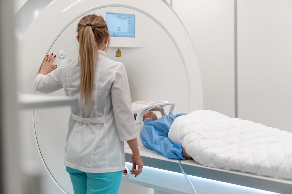
Your MRI Appointment: A Step-by-Step Guide to What to ExpectKnowing what will happen on the day can make a world of difference, turning an anxious wait into a calm and managed experience. Let's walk through it together, step by step, so you feel completely prepared. Step 1: Arrival and Check-In When you arrive at the MRI unit, you'll check in with the receptionist. They'll ask you to confirm your details and then hand you a final, very thorough safety questionnaire. Don't be alarmed by the number of questions; this is a routine part of the process for every single patient. It's designed to be the ultimate safety net. Take your time, read each question carefully, and answer honestly. This is your chance to flag your implant again, even if you’ve already done so ten times. Step 2: The Consultation with the Radiographer A radiographer will come and collect you from the waiting area and take you to a private space. Think of them as your personal guide for the scan. Their number one priority is your safety and comfort. They will sit with you and go through your questionnaire, paying special attention to your implant. This is the moment to present your patient implant card or the documentation you’ve gathered. The radiographer will be genuinely grateful that you've come prepared. They will use the manufacturer and model number to look up your specific device in their safety database. This tells them the exact 'conditions' for your scan – the power settings, the scan duration, and any other parameters they need to follow to the letter. This isn't just paperwork for them; it's the professional checklist that guarantees your safety. Feel free to ask them any questions you have. No question is too small or silly. Step 3: Getting Changed and Final Checks You'll be shown to a private changing room and asked to change into a hospital gown. You will need to remove absolutely everything else – watches, all jewellery (including wedding rings and piercings), glasses, hearing aids, and any removable dental work. You'll be given a secure locker for your valuables. This might seem extreme, but it's vital. Even tiny metallic threads in socks or clasps on underwear can heat up or distort the images, so it’s a standard rule for everyone. Step 4: Entering the Scan Room and Getting Comfortable The radiographer will lead you into the scan room. It’s usually a large, bright room dominated by the MRI scanner, which looks like a large white doughnut. They will help you onto the cushioned motorised bed that slides into the scanner. Their goal is to make you as comfortable as possible, using pillows and foam pads to support your body. They will then place a piece of equipment called a 'coil' on or around the part of your body being scanned. It might look like a plastic cage or a blanket, but it's essentially an antenna that helps get the best quality picture. You'll be given earplugs and a set of headphones. This is very important, as the scanner is incredibly loud. Here's a great tip: many departments allow you to listen to music! You can often connect to Spotify or listen to various radio stations. Ask about this beforehand. Listening to your favourite music can be a fantastic way to distract yourself and make the time pass quickly. Finally, they will give you a rubber squeeze-ball or a buzzer. This is your direct line to them. If you feel any discomfort, anxiety, or just need to stop, you squeeze it, and they will immediately stop the scan and talk to you over the intercom. You are in control at all times. Step 5: During the Scan The radiographer will move to the control room next door, where they can see and hear you throughout the entire scan. They will let you know over the intercom that they're about to start. The bed will gently slide into the scanner. For most scans, your head will remain outside the tunnel unless, of course, it's your head or neck being scanned. Then the noises will begin. It's a series of very loud clanking, buzzing, and rhythmic thumping sounds. It can be a bit startling at first, but it helps to think of it as the machine's 'work song'. Try to close your eyes, focus on your breathing, and tune into your music. Your only job is to lie as still as you possibly can. The scan is performed in sequences, so you'll have the loud noise for a few minutes, followed by a brief, quiet pause. The radiographer will often update you, saying things like "That was the first set, you're doing great, just three more to go." Step 6: All Finished Once the last image is taken, the bed will slide back out. The radiographer will come in and help you up. You might feel a little stiff from staying still, but that's it. You can head back to the changing room, get dressed, and collect your belongings. Usually, you can leave straight away and go about your day as normal. The images will be reviewed by a specialist radiologist, and a report will be sent to your doctor in a few days. You’ve done it! 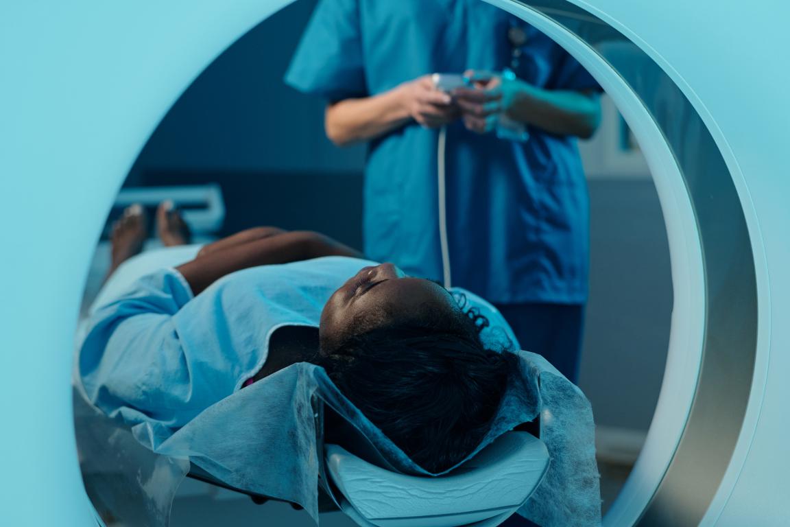
From Information to Peace of Mind: A Simple Step for Your SafetyWe've covered a lot of ground, from the science of magnets to the practical steps of a hospital appointment. The single common thread that runs through it all is the critical importance of having accurate and accessible information about your implant. Your safety during an MRI depends entirely on the medical team knowing the exact make, model, and manufacturer of your device. Without it, they simply cannot proceed. We know that finding this information can be a stressful process. It often involves digging through old paperwork, making phone calls to busy hospital departments, and waiting for records to be sent – all while you're likely already worried about your health. Imagine arriving for your appointment, having taken time off work and arranged transport, only to have it cancelled because you couldn't find that one crucial piece of paper. It's a frustrating and preventable situation. This is where planning ahead can provide enormous peace of mind. Having your vital implant details stored on a single, clear, and durable card that you can carry with you at all times removes that last-minute panic. It transforms a moment of stress into a simple, confident action. You can just hand over the card, knowing that the radiographer has everything they need to keep you safe. It puts you in control of your own health information and helps ensure your medical journey is as smooth and stress-free as possible. Taking this one simple step can make all the difference. Learn more about how you can stay prepared with our complete guide to Metal Implant ID Cards.
© 2024 The Card Project Uk Ltd
VAT: 453 2087 06
|